
Mouse Secondary Antibodies

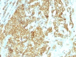
Biotium Glucose Regulated Protein 94 (GRP94)(HSP90B1/1192), 0.2mg/mL
Recognizes a protein of 94 kDa, which is identified as the glucose-regulated protein 94 (grp94) and also tumor rejection antigen (gp96). Grp94 shows a high degree of sequence homology with the heat shock protein 90 (hsp90). This MAb is highly specific to grp94 and shows minimal cross-reaction with other members of the HSP90 family. Grp s are a class of proteins unresponsive to heat shock and are induced by glucose deprivation. Grp94 has been briefly studied as a prognostic factor in breast cancer.Primary antibodies are available purified, or with a selection of fluorescent CF Dyes and other labels. CF Dyes offer exceptional brightness and photostability. Note: Conjugates of blue fluorescent dyes like CF405S and CF405M are not recommended for detecting low abundance targets, because blue dyes have lower fluorescence and can give higher non-specific background than other dye colors.


Biotium Prostate Specific Antigen (PSA)(KLK3/1248), CF740 conjugate, 0.1mg/mL
Recognizes a single protein of 33-34 kDa, identified as the prostate specific antigen (PSA). This MAb is highly specific to PSA and stains prostatic secretory and ductal epithelium in both normal and neoplastic tissues. PSA is a chymotrypsin-like serine protease (kallikrein family) exclusively produced by the prostate epithelium, and abundant in seminal fluid. PSA can be detected in the sera of patients with prostatic carcinoma. It is predominantly complexed to a liver-derived serine protease inhibitor, alpha-1-antichymotrypsin (ACT). A higher proportion of serum PSA is complexed to ACT in prostate cancer than in benign prostate hyperplasia. This MAb makes an excellent pair with MAb A67-B/E3 for PSA tests.
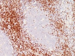
Biotium CD6(C6/372 + 3F7B5), CF740 conjugate, 0.1mg/mL
CD6 is a type I transmembrane glycoprotein that contains a 24-amino acid signal sequence, three extracellular scavenger receptor cysteine-rich (SRCR) domains, a membrane-spanning domain and a 44-amino acid cytoplasmic domain. The CD6 glycoprotein is tyrosine phosphorylated during TCR-mediated T cell activation. CD6 shows significant homology to CD5. CD6 is present on mature thymocytes, peripheral T cells and a subset of B cells. Antibodies to CD6 are used to deplete T cells from bone marrow transplants to prevent graft versus host disease.Primary antibodies are available purified, or with a selection of fluorescent CF Dyes and other labels. CF Dyes offer exceptional brightness and photostability. Note: Conjugates of blue fluorescent dyes like CF405S and CF405M are not recommended for detecting low abundance targets, because blue dyes have lower fluorescence and can give higher non-specific background than other dye colors.
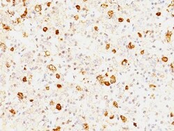
Biotium FSH beta(FSHb/1062), CF568 conjugate, 0.1mg/mL
This MAb reacts with a protein of 22 kDa, identified as beta sub-unit of FSH. It does not cross react with the alpha sub-unit. Follicle stimulating hormone (FSH) is a hormone synthesized and secreted by gonadotrophs in the anterior pituitary gland. In the ovary, FSH stimulates the growth of immature Graafian follicles to maturation. As the follicle grows, it releases inhibin, which deactivates the FSH production. In men, FSH enhances the production of androgen-binding protein by the Sertoli cells of the testis and is critical for spermatogenesis. FSH and LH act synergistically in reproduction. FSH is a useful marker in the classification of pituitary tumors and the study of pituitary disease.Primary antibodies are available purified, or with a selection of fluorescent CF Dyes and other labels. CF Dyes offer exceptional brightness and photostability. Note: Conjugates of blue fluorescent dyes like CF405S and CF405M are not recommended for detecting low abundance targets, b
Biotium Prostate Specific Antigen (PSA)(1A7), CF740 conjugate, 0.1mg/mL
This antibody recognizes a single protein of 33-34 kDa, identified as the prostate specific antigen (PSA). This MAb is highly specific to PSA and stains prostatic secretory and ductal epithelium in both normal and neoplastic tissues. PSA is a chymotrypsin-like serine protease (kallikrein family) exclusively produced by the prostate epithelium, and abundant in seminal fluid. PSA can be detected in the sera of patients with prostatic carcinoma. It is predominantly complexed to a liver-derived serine protease inhibitor, alpha-1-antichymotrypsin (ACT). A higher proportion of serum PSA is complexed to ACT in prostate cancer than in benign prostate hyperplasia. This MAb makes an excellent pair with MAb A67-B/E3 for PSA tests.
Biotium CD46(169-1-E4.3), CF740 conjugate, 0.1mg/mL
Recognizes a protein of 52 kDa-58 kDa, identified as CD46 (also known as membrane cofactor protein, MCP). Its epitope is located on the four immunogenic short consensus repeat (SCR) domains at N-terminus of the protein. CD46 exists as many isoforms in a variety of tissues. It is strongly expressed on salivary gland ducts and kidney ducts, moderately on lymphocytes and endothelium, and weakly on interstitial tissues and muscle cells, but not on erythrocytes. CD46 functions as a C3b/C4b-binding glycoprotein that inhibits complement activation on host cells. It also serves as a measles virus receptor, an adherence factor for group A Streptococcus pyogenes, and a cellular pilus receptor for pathogenic Neisseria.Primary antibodies are available purified, or with a selection of fluorescent CF Dyes and other labels. CF Dyes offer exceptional brightness and photostability. Note: Conjugates of blue fluorescent dyes like CF405S and CF405M are not recommended for detecting low abun
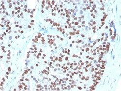
Biotium NKX3.1 (Metastatic Prostate Adenocarcinoma Marker)(NKX3.1/3348), CF568 conjugate, 0.1mg/mL
NKX3. 1 is a prostate specific gene encoding a transcription factor that plays an important role in normal prostate development and carcinogenesis. It is a prostatic tumor suppressor gene located on chromosome 8p21. 2, which frequently undergoes a loss of heterozygosity. NKX3. 1 expression is highly restricted in prostate epithelial cells and therefore can be used as a diagnostic biomarker for prostate cancer and other metastatic lesions of prostatic origin. Furthermore, NKX3. 1 shows better sensitivity than Prostate Specific Antigen (PSA) for identifying metastatic prostatic adenocarcinoma. This suggests that immunohistochemical staining of NKX3. 1, along with other prostate-restricted markers, may be valuable for the definitive determination of prostatic origin in poorly differentiated metastatic carcinomas. Primary antibodies are available purified, or with a selection of fluorescent CF Dyes and other labels. CF Dyes offer exceptional brightness and photostability. Note
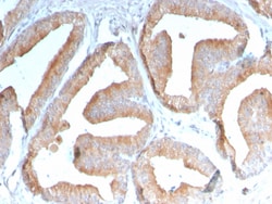
Biotium ICOS-L / ICOS Ligand / B7RP-1 (Immuno-Oncology Target)(ICOSL/3111), CF488A conjugate, 0.1mg/mL
This MAb recognizes a protein of 36 kDa, identified as ICOS-L. It is a ligand for the ICOS receptor that initiates T and B cell proliferation and cytokine secretion. ICOS-L interactions play an essential role in T cell-dependent B cell activation in peripheral lymphoid organs such as spleen and lymph nodes. ICOS-L protein is present in myeloid leukocytes, and by Northern blot there are 2. 4, 3. 0, and 7. 0 kb transcripts in brain, heart, kidney, and liver, with lower expression in colon and thymus, and a 1. 1 kb transcript in leukocytes. Tumor necrosis factor alpha (TNF), granulocyte-macrophage colony-stimulating factor (GM-CSF) and interleukin-4 (IL-4) enhance its expression. LPS-induced up-regulation of ICOS-L is dependent on the MyD88-dependent signaling pathway. Primary antibodies are available purified, or with a selection of fluorescent CF Dyes and other labels. CF Dyes offer exceptional brightness and photostability. Note: Conjugates of blue fluorescent dyes like CF
Revvity Health Sciences Inc HTRF Human Total ULK1 Detection Kit, 500 Assay Points
HTRF Human Total ULK1 Detection Kit, 500 Assay Points

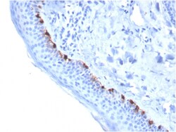
Biotium Tyrosinase-Related Protein-1 (TYRP-1) (Melanoma Marker)(TYRP1/3284), CF568 conjugate, 0.1mg/mL
Reacts with a 75 kDa melanocyte-specific gene product, identified as Tyrosinase-related protein-1 (TRP-1). It is involved in melanin synthesis. TRP1 is present on the melanosomal membranes of melanoma, normal melanocytes and nevi. Recent evidence suggests that TRP-1 is involved in maintaining stability of tyrosinase protein and modulating its catalytic activity. TRP-1 is also involved in maintenance of melanosome ultrastructure and affects melanocyte proliferation and cell death. Primary antibodies are available purified, or with a selection of fluorescent CF Dyes and other labels. CF Dyes offer exceptional brightness and photostability. Note: Conjugates of blue fluorescent dyes like CF405S and CF405M are not recommended for detecting low abundance targets, because blue dyes have lower fluorescence and can give higher non-specific background than other dye colors.
Biotium NOX4 / NADPH Oxidase 4 (NOX4/1245), CF568 conjugate, 0.1mg/mL
The superoxide-generating NADPH oxidase includes a membrane-bound flavocytochrome containing two subunits, gp91-phox and p22-phox, and the cytosolic proteins p47-phox and p67-phox. During activation of the NADPH oxidase, p47-phox and p67-phox migrate to the plasma membrane where they associate with the flavocytochrome, cytochrome b558, to form the active enzyme complex. The p22 and gp91-phox subunits also function as surface O2 sensors that initiate cellular signaling in response to hypoxic conditions. NOX4 is a renal gp91-phox homolog highly expressed at the site of erythropoietin production in the proximal convoluted tubule epithelial cells of the renal cortex. It is also expressed in fetal tissues, placenta, glioblastoma and vascular cells.Primary antibodies are available purified, or with a selection of fluorescent CF Dyes and other labels. CF Dyes offer exceptional brightness and photostability. Note: Conjugates of blue fluorescent dyes like CF405S and CF405M are no
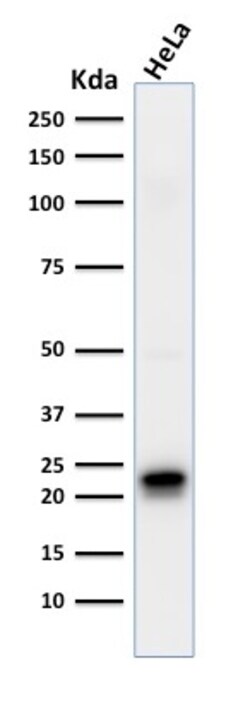
Biotium Ferritin, Heavy Chain (FTH) (Microglia Marker)(FTH/2081), CF488A conjugate, 0.1mg/mL
Mammalian ferritins consist of 24 subunits made up of 2 types of poly-peptide chains, ferritin heavy chain and ferritin light chain, which each have unique functions. Ferritin heavy chains catalyze the first step in iron storage, the oxidationof FeI(II), whereas ferritin light chains promote the nucleation of ferrihydrite, enabling storage of Fe(III). The most prominent role of mammalian ferritins is to provide iron-buffering capacity to cells. In addition to iron buffering, heavy chain ferritin is also involved in the regulation of thymidine biosynthesis via increased expression of cytoplasmic serine hydroxymethyltransferase, which is a limiting factor in thymidylate synthesis in MCF-7 cells. Light chain ferritin is involved in cataracts by at least two mechanisms: hereditary hyperferritinemia cataract syndrome, in which light chain ferritin is overexpressed; and oxidative stress, an important factor in the development of aging-related cataracts. Primary antibodies are available
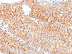
Biotium PAI-RBP1 / SERBP1 / SERPINE1(SERBP1/3491), 1mg/mL
PAI-RBP1 (plasminogen activator inhibitor 1 RNA-binding protein), also known as SERBP1 (SERPINE1 mRNA-binding protein 1), CGI-55, CHD3IP (chromodomain helicase DNA binding protein 3 interacting protein), HABP4L or PAI-RBP1, is a membrane-associated protein that localizes to the nucleus, the perinuclear region of the cytoplasm and the plasma membrane. PAI-RBP1 is believed to play a role in the regulation of mRNA stability, as it specifically binds to the CRS (cyclic nucleotide-responsive sequence) motif of the PAI-1 mRNA and acts to stabilize the mRNA and regulate its expression. In addition, PAI-RBP1 interacts with Mi2- and may be involved in chromatin remodeling. PAI-RBP1 also interacts with PGRMC1 and participates in the transduction of Progesterone's antiapoptotic action in ovarian cell types. The gene encoding PAI-RBP1 is overexpressed in ovarian cancer, suggesting a possible role for PAI-RBP1 in tumorigenesis and tumor metastasis. Primary antibodies are available purifie
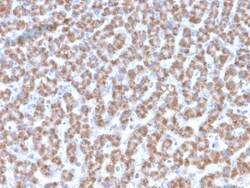
Biotium Prohibitin (Mitochondrial Marker)(PHB/3193), 0.2mg/mL
Recognizes a protein of 30 kDa which is identified as Prohibitin, an evolutionarily conserved protein with homologues found in yeast to man. It is located in the inner membrane of mitochondria. Although prohibitin mRNA and protein expression occurs throughout the cell cycle, maximum levels are detected during the G1/S phase transition and minimum levels are seen in S phase and the G2/mitosis boundary. Prohibitin is located exclusively in the mitochondria with the highest concentration on the inner membrane. Prohibitin is an ideal mitochondrial marker. It shows antiproliferative activity and has been proposed to play a role in normal cell cycle regulation, replicative senescence, cellular immortalization, and tumor suppression. Primary antibodies are available purified, or with a selection of fluorescent CF Dyes and other labels. CF Dyes offer exceptional brightness and photostability. Note: Conjugates of blue fluorescent dyes like CF405S and CF405M are not recommended fo
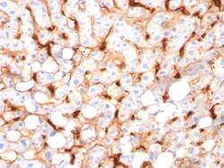
Biotium C1QB / Complement C1q B-Chain(C1QB/2966), CF740 conjugate, 0.1mg/mL
Complement C1q B Chain (C1qb) is a subunit of C1q. C1q is composed of 6 polypeptide chains each of C1qa, C1qb, and C1qc. C1q is a component of the classical complement pathway, and thereby plays an important role in the immune response. Each C1q subunit contains an N-terminal collagen-like region and a C-terminal C1q globular domain. The presence of receptors for C1q on effector cells modulates its activity, which may be antibody-dependent or independent. Macrophages are the primary source of C1q, while anti-inflammatory drugs as well as cytokines differentially regulate expression of the mRNA as well as the protein. C1q deficiency is associated with lupus erythematosus and glomerulonephritis. Primary antibodies are available purified, or with a selection of fluorescent CF Dyes and other labels. CF Dyes offer exceptional brightness and photostability. Note: Conjugates of blue fluorescent dyes like CF405S and CF405M are not recommended for detecting low abundance targets,